
Anatomy Of The Teres Minor Muscle —
Gross anatomy. The muscle originates from the dorsal surface of the inferior angle of the scapula and inserts on the medial lip of the intertubercular groove of the humerus. It is related to the latissimus dorsi muscle which wraps around the lower border of teres major. The tendon of the teres major lies posterior to the tendon of the.
Anatomy of the Teres Major —
Teres Major Anatomy Palpation. Palpating the teres major is an essential technique for clinicians to assess its condition and identify potential issues. To palpate the teres major, the patient should be in a seated or lying position, with the arm slightly adducted and medially rotated.
Teres Major Sehne Nehru Memorial
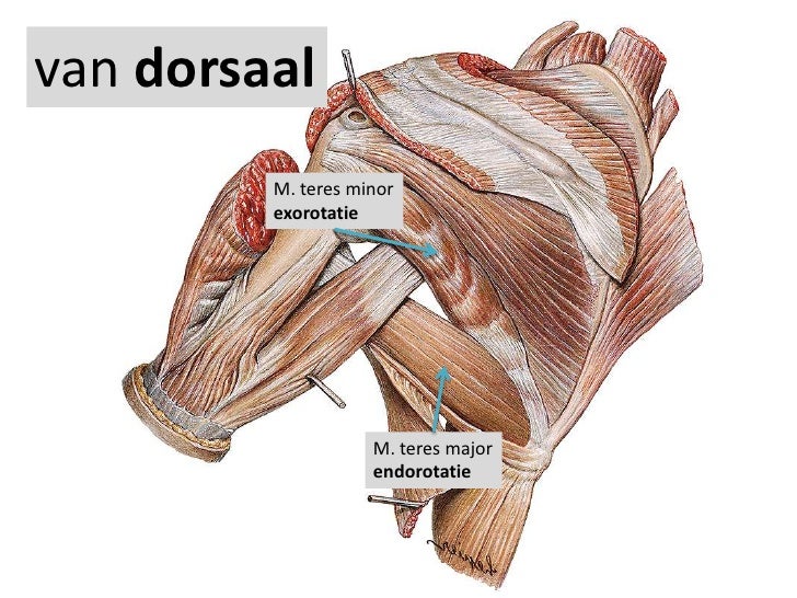
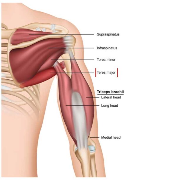
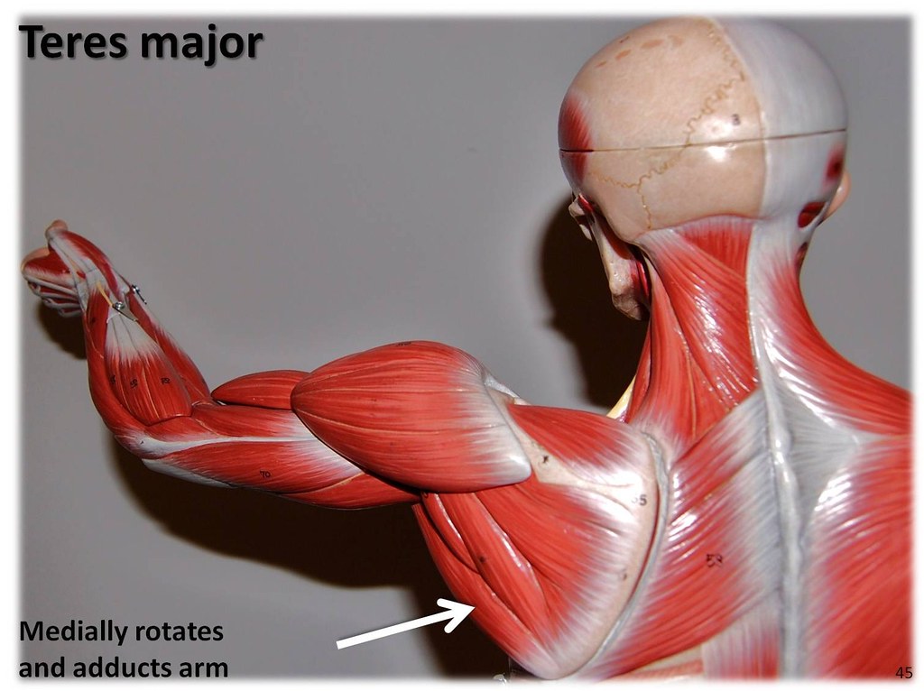
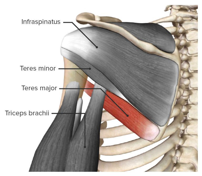
The teres major muscle is found in the shoulder region. It is a thick, fusiform type of skeletal muscle. It is located: - anterior (deep) to the long head of triceps brachii muscle; - posterior (superficial) to the scapula, coracobrachialis muscle; - superior to the latissimus dorsi muscle; - inferior to the teres minor and infraspinatus muscles.
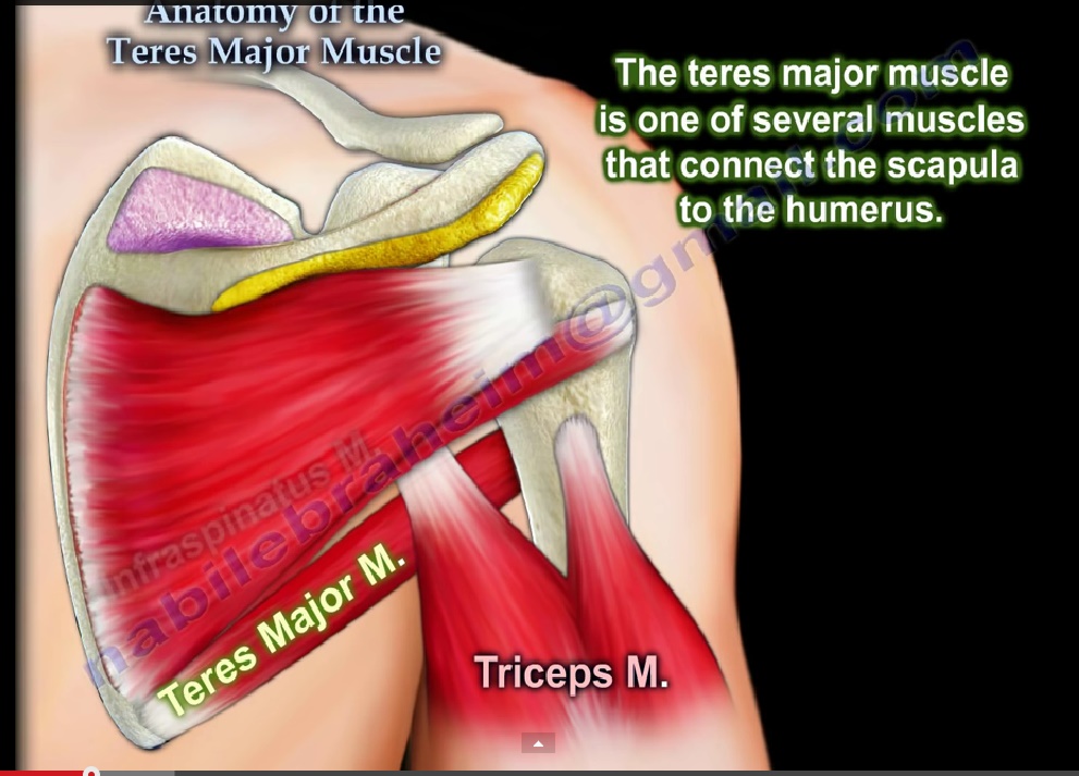
Anatomy Of The Teres Major Muscle —
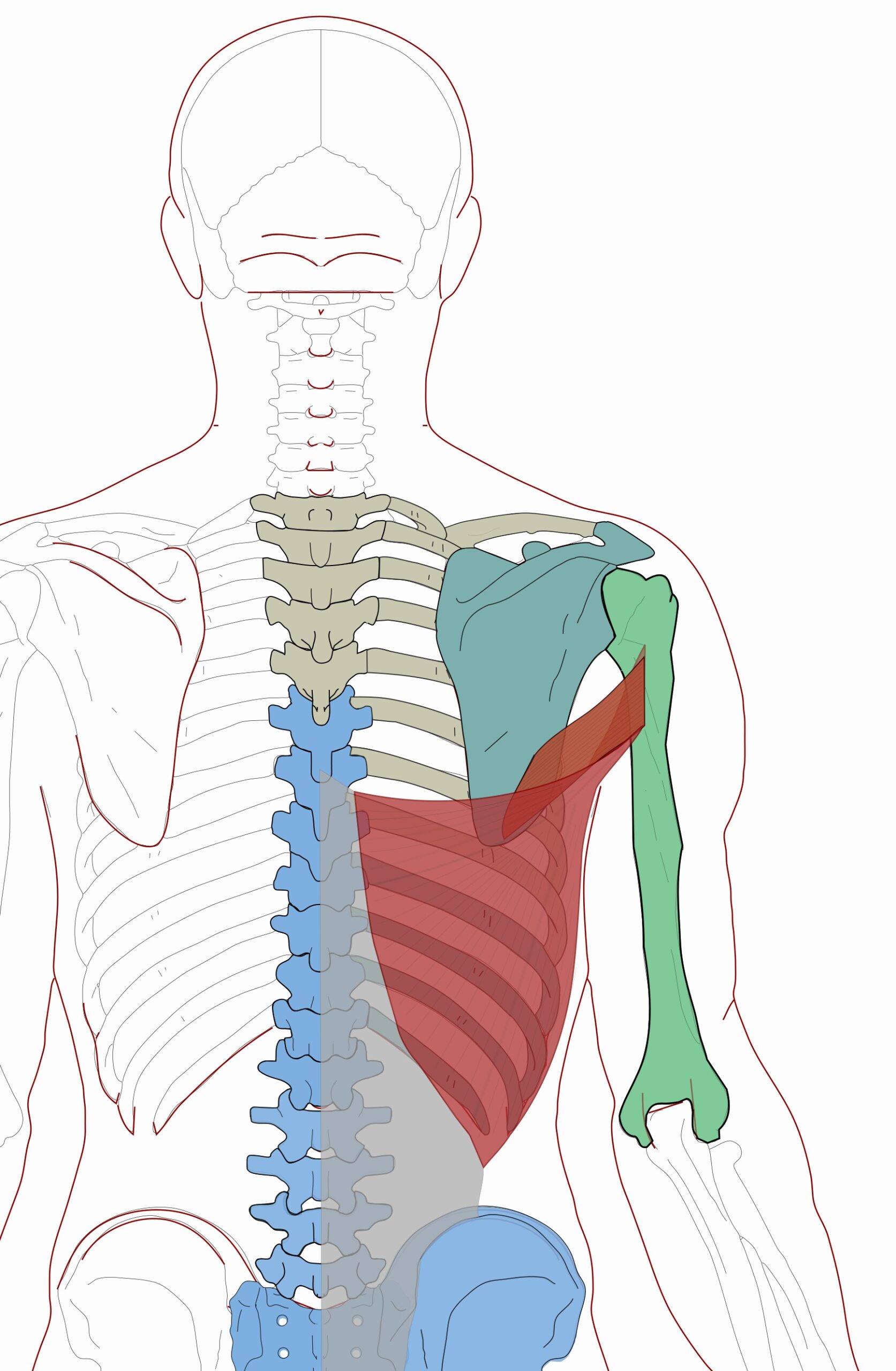
The teres major is a thick but flattened, rectangular muscle that extends from the inferior posterior scapula to the medial lip of the intertubercular groove of the humerus.[1][2] It functions synergistically with the latissimus dorsi to extend, adduct, and internally rotate the humerus.[3] Although the latissimus dorsi and teres major muscles often function in conjunction with one another.

The Teres Major Muscle
Teres major muscle (Musculus teres major) The teres major is a thick muscle of the shoulder joint. It spans from the inferior aspect of the scapula to the proximal part of the humeral shaft. Unlike the teres minor, the teres major muscle does not attach to the capsule of the glenohumeral joint. Thus it is not regarded as part of the rotator.
Teres Major Origin, Insertion, Action The Wellness Digest
Dr. Ebraheim's educational animated video describes the anatomy of the Teres Major muscle.Origin & insertion: the teres major muscle arises from the dorsal (.

Anatomy Of The Teres Major Orthopaedicprinciples Com Gambaran
The teres major muscle is a muscle of the upper limb.It attaches to the scapula and the humerus and is one of the seven scapulohumeral muscles.It is a thick but somewhat flattened muscle. The teres major muscle (from Latin teres, meaning "rounded") is positioned above the latissimus dorsi muscle and assists in the extension and medial rotation of the humerus.
Teres major Muscles of the Upper Extremity Visual Atlas,… Flickr
If your teres major is overactive/short, do the following: Reduce your training volume on teres major exercises and lat exercises. If your overhead range of motion is limited, avoid vertical push exercises like the overhead press. Do high-incline push exercises instead (e.g. shoulder press on 60-75° incline).

teres major bodybuilding Google Search
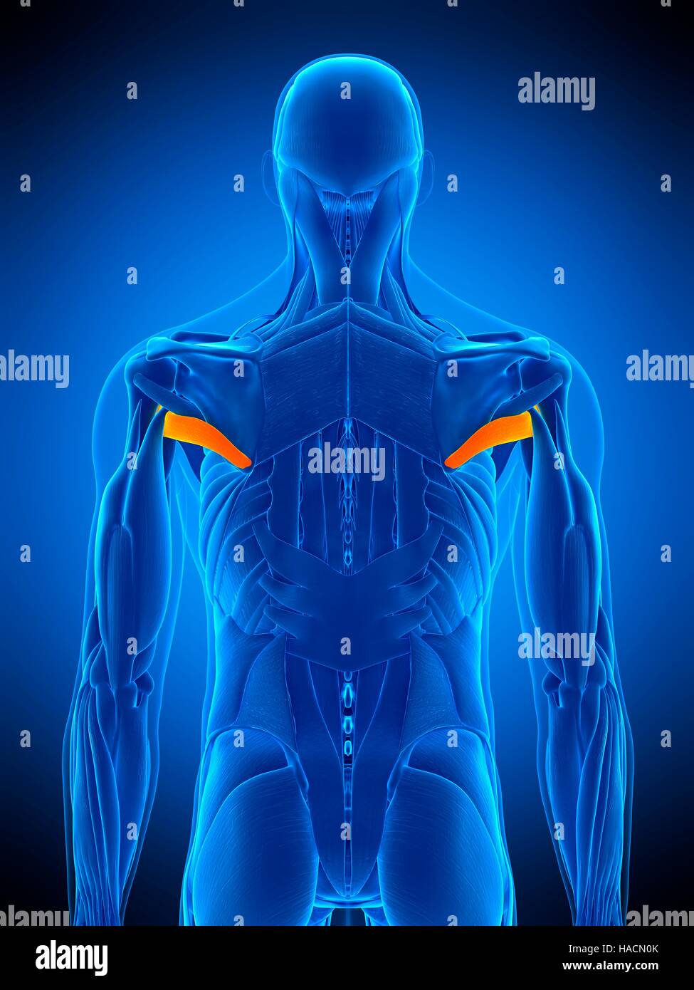
Teres major. The teres major muscle is one of the six muscles within the scapulohumeral muscle group. The muscle is located on the underside of the upper arm, in the area between the shoulder and.

15 Teres Major And Teres Minor Strengthening Exercises SET FOR SET
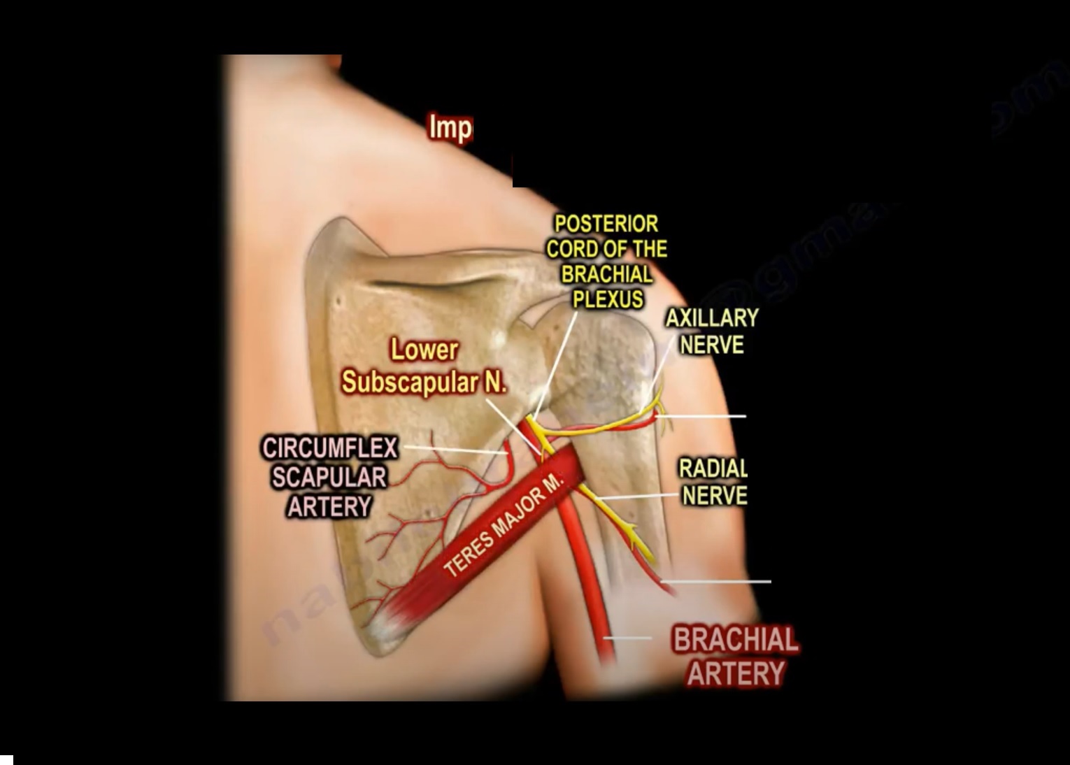
Origin: Posterior aspect of the inferior angle of the scapulaInsertion: Medial lip of the intertubercular sulcus of the humerusArtery: Subscapular and circumflex scapular arteriesNerve: Lower subscapular nerve (segmental levels C5 and C6)Action: Internal rotation of the humerus Description: The Teres major is a thick but somewhat flattened muscle, which arises from the oval area on the dorsal.

Latissimus dorsi Musculus teres major Musculus teres minor Muskelherkunft und Insertionsanatomie
The teres major is a thick but flattened, rectangular muscle that extends from the inferior posterior scapula to the medial lip of the intertubercular groove of the humerus. It functions synergistically with the latissimus dorsi to extend, adduct, and internally rotate the humerus. Although the lati.
Shoulder Joint Anatomy Concise Medical Knowledge
In this video we go over the anatomy facts of the teres major muscle: origin, insertion, innervation and function. Test yourself in our arm and shoulder musc.
Teres Major Functional Anatomy Integrative Works
ANATOMY. Teres Major. Origin. Dorsal surface of inferior angle of scapula. Insertion. Medial lip of intertubercular groove of humerus. Action. Adducts and medially rotates arm. Innervation.

Teres Major Muscle Origin, Insertion & Action Human Anatomy Kenhub YouTube
Teres Major injuries result in pain and difficulty with activities that require sideways or backwards movements with the arm. Isolated tears of the teres major are quite uncommon, but may occur in baseball or cricket players, especially pitchers and bowlers.; The main symptom of a teres major tear is a sudden sharp pain in the shoulder, upper arm and armpit.
Teres major muscle hires stock photography and images Alamy
Dorsal surface of inferior angle of scapula. Insertion. Medial lip of intertubercular groove of humerus. Action. Adducts and medially rotates arm. Innervation. Lower subscapular nerve (C6 and C7) (C6, C7) Arterial Supply. Subscapular and circumflex scapular arteries.
muscle connection anatomy
⭐ Teres Major Muscle Anatomy ⭐💪 Origin: Inferior angle of the medial border of the scapula.💪 Insertion: Medial crest of the intertubercular groove (also kn.